New algorithms help interpret vision loss from digital images
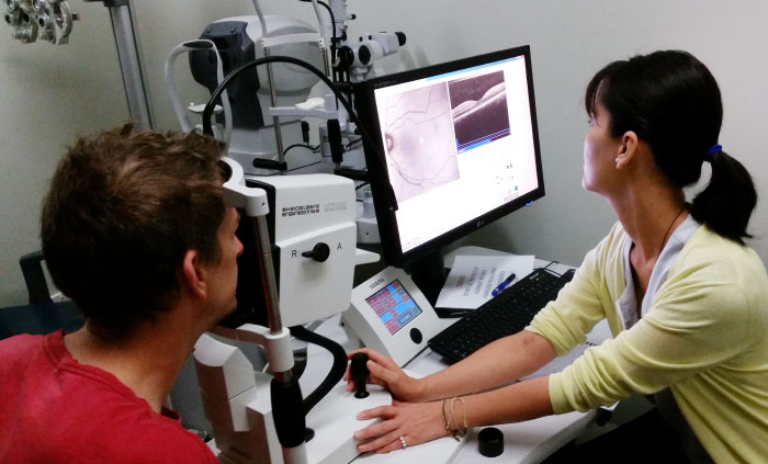
Technology that can take a three-dimensional image of the inside of the human eye is revolutionising eye care. These images are especially suited to detecting and monitoring two of the most common causes of progressive vision loss: glaucoma and age-related macular degeneration.
Advances in computing power and LCD screen technology have both contributed to the adoption of digital imaging in eye care. However, the brain plays such a powerful role in generating our sense of sight that damage to the light-sensing retina does not necessarily result in a one-to-one corresponding loss of vision.
That means retinal images — no matter how sophisticated — require interpretation.
To correct this diagnostic gap, several manufacturers of retinal imaging machines have turned to Andrew Turpin at the University of Melbourne’s School of Computing and Information Systems for solutions.
In the case of glaucoma, Professor Turpin worked with German company Heidelberg Engineering to develop a software fix that better translates 3-D retinal images into patient-relevant information.
Professor Turpin’s approach was developed based on his study of the patterns that optic nerve axons create as they grow during foetal development, particularly the tendency of these axons to take the shortest path across the back of the retina to exit towards the brain.
The relationship between damage to the retina and impacts on vision have been overly simplified. Without the software correction, the digital imaging technology may underestimate the rate with which glaucoma patients progressively lose vision.
Professor Andrew Turpin
Heidelberg Engineering has since begun to incorporate Professor Turpin’s biologically compatible software into its imaging machines, improving diagnostics and eye care for those affected by glaucoma.